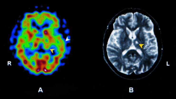
Brain Single Photon Emission Computed Tomography (SPECT) is a sophisticated imaging method that utilizes radioactive tracers to examine and visualize brain function. This nuclear medicine technique allows health professionals to evaluate the physiological processes within the brain, effectively making it possible to gain a snapshot of regional cerebral blood flow and metabolic activity.
Unlike traditional MRI or CT scans, which primarily focus on structural anatomy, SPECT provides critical information about how the brain is working in real-time, thus offering insights into functional abnormalities that may not be apparent from structural imaging alone.
The SPECT imaging process involves injecting a small amount of a radioactive agent, followed by waiting for the agent to circulate and accumulate in the brain, where it emits gamma rays. A gamma camera detects these rays and creates 3D images that represent brain functionality. This imaging technique is invaluable in a variety of fields, including psychiatry, neurology, and cardiovascular medicine.
Understanding the operational dynamics of the brain can help in diagnosing conditions such as Alzheimer’s disease, epilepsy, and depression, making SPECT an indispensable tool in modern medicine.
Distinctive Features ─ How SPECT Differs from Other Imaging Techniques
One of the most striking differences between SPECT and other imaging modalities, such as MRI and CT, is the focus on functional rather than structural mapping. While MRI and CT provide detailed images of brain anatomy, SPECT highlights the blood flow and metabolic processes taking place within various brain regions.
This functional information is critical in understanding disorders that exhibit no overt structural changes, such as certain mood disorders or neurodegenerative diseases. SPECT is particularly sensitive to changes in cerebral blood flow and, thus, provides deeper clinical insights during the early stages of disease progression.
Moreover, SPECT scans can be performed relatively quickly and may require less complex equipment compared to MRI, making it more accessible to a range of healthcare facilities. Additionally, while MRI offers superior resolution for detecting structural abnormalities such as tumors or strokes, SPECT is more effective in revealing the brain’s functional metabolism, allowing for a comprehensive understanding of cognitive and mood variations in patients.
Real-life Applications ─ When and Why is Brain SPECT Used?
Brain SPECT imaging is employed in various clinical scenarios that require operational insights into brain functions. It is particularly useful in diagnosing neurological conditions such as stroke, epilepsy, and head trauma, where understanding the brain’s metabolic patterns can guide therapeutic decisions. In psychiatry, SPECT can illuminate the underlying processes of disorders such as schizophrenia, bipolar disorder, and major depressive disorder, thereby assisting practitioners in tailoring personalized treatment plans.
Furthermore, SPECT is instrumental in monitoring responses to therapeutic interventions, such as how a patient’s brain activity evolves following pharmacological treatment or psychotherapy. This dynamic aspect of SPECT allows for adjustments in treatment regimens based on quantitative data rather than solely relying on subjective reports and behavioral assessments. Conditions like Alzheimer’s disease may benefit significantly from SPECT, as it can elucidate disruptions in brain metabolism, facilitating early detection and management strategies that might improve quality of life.
Unlocking the Mysteries of Brain Function ─ Understanding the Insights from SPECT
Mapping Brain Activity ─ What Does Your Brain SPECT Scan Reveal?
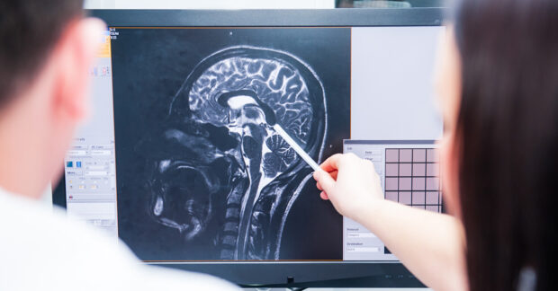
The output of a brain SPECT scan is a powerful visual representation of the brain’s functional status. Each region of the brain has specific responsibilities from those controlling motor functions to areas associated with cognition, emotions, and memory.
By assessing regional cerebral blood flow, SPECT can reveal patterns of activation or hypoactivity linked to different neurological and psychological states. For example, elevated activity in certain areas may correlate with mania in bipolar disorder, whereas diminished activity may indicate depressive states.
These functional patterns help clinicians not only diagnose existing conditions but also predict the course of the illness by identifying underlying neural dysfunctions that manifest as behavioral or cognitive deficits.
Each patient may exhibit unique patterns, providing essential insights for personalized treatment approaches. In particular, understanding these patterns can improve therapeutic outcomes by tailoring interventions that support the specific areas requiring rehabilitation or enhancement in cognitive function.
Insights into Mental Health ─ Can SPECT Help Diagnose Conditions?

The realm of mental health is complex, often requiring nuanced approaches to diagnosis and treatment. Brain SPECT imaging plays a pivotal role in depathologizing mental health disorders by illustrating the neurobiological substrates underlying psychiatric conditions. Studies have shown significant deviations in blood flow patterns in conditions such as major depressive disorder, obsessive-compulsive disorder (OCD), and post-traumatic stress disorder (PTSD), which are typically elusive when relying solely on behavioral assessments.
Through brain SPECT, healthcare providers can establish more accurate diagnoses, paving the way for targeted therapies. For instance, SPECT can indicate which neurotransmitter systems may be dysfunctional, facilitating the decision to use specific medication types. This capability to provide quantifiable data enhances the overall therapeutic strategy and allows for a more objective evaluation of treatment effectiveness over time, ensuring that the patient’s journey toward recovery is both transparent and measurable.
Exploring Neurobiological Patterns ─ What Your Scan Says About You
Each individual’s brain is unique, akin to a fingerprint, and brain SPECT imaging can highlight these individual differences in neurobiological activity. The qualitative analysis of SPECT reveals how various brain networks interact and perform under different stress and stimulus conditions information that can be pivotal in understanding a person’s cognitive and emotional landscape. Patients can see how their brain functions align or diverge from normative data, providing significant insights into their behaviors, choices, and emotional responses.
Moreover, SPECT imaging can serve as a tool for self-reflection and personal development. By comprehensively understanding one’s brain functions, patients can engage in more informed discussions with mental health professionals about their conditions, accelerating the treatment process. Recognizing these patterns empowers individuals to actively participate in their care, fostering a greater sense of agency and cooperation in therapeutic relationships.
Brain SPECT and Holistic Health ─ Bridging the Gap Between Mind and Body
The Role of Brain SPECT in Personalized Treatment Plans
Personalized medicine emphasizes tailored treatment strategies that consider individual variances in genetics, lifestyle, and environmental contexts. Brain SPECT imaging provides critical data that dovetail with this approach, effectively guiding clinicians in developing targeted treatment protocols. By incorporating functional data into treatment planning, healthcare professionals can select therapies from pharmaceutical treatments to behavioral interventions that correspond closely with the patient’s unique neurobiology.
This holistic approach transcends traditional one-size-fits-all treatments, promoting strategies that are more likely to yield successful outcomes. For instance, if SPECT scanning reveals specific hypoactive regions associated with cognitive deficits, clinicians might introduce cognitive training exercises that effectively engage those areas, thereby stimulating neuroplasticity and helping to improve cognitive function over time.
Moreover, therapies may be adjusted based on ongoing SPECT assessments, allowing for real-time tracking of brain function improvements or regressions, which is particularly significant in chronic conditions. This dynamic aspect enriches the therapeutic alliance, helping both patients and providers co-create treatment strategies to better engage different neural networks conducive to recovery.
Integrating SPECT Findings into Wellness Strategies
Utilizing the insights gained from brain SPECT assessments enriches overall wellness strategies across various domains, including mental health, physical health, and lifestyle optimization. Understanding how neural patterns correlate with mental performance can inform decisions about diet, sleep, exercise, and stress management. For instance, if a SPECT scan indicates decreased activity in specific areas during stress, practitioners may recommend practices such as mindfulness, yoga, or brain-training exercises designed to foster resilience and enhance brain functionality.
Additionally, collaborative approaches involving nutrition, exercise, and cognitive behavioral strategies can be woven into a comprehensive treatment plan. Knowledge gleaned from a SPECT scan can provide crucial motivation for patients to adopt healthier lifestyle choices, strengthening their psychological commitment to the treatment process.
By aligning therapeutic efforts with functional brain data, patients can experience a more profound sense of engagement and mastery over their health.
The Future of Brain Imaging ─ A Look at Emerging Technologies
The landscape of brain imaging is continuously evolving, with advancements in technology promising exciting prospects for functional imaging methodologies like SPECT. Innovations in the precision of imaging agents, alongside advanced computational techniques, are enhancing the resolution and sensitivity of SPECT, thereby facilitating an even richer understanding of neural operations. Future developments might include integrated imaging approaches that combine the strengths of SPECT with other modalities such as functional MRI (fMRI) or advanced electrophysiological assessments.
Further research into artificial intelligence and machine learning algorithms could revolutionize the interpretation and analysis of SPECT data, leading to more precise diagnoses and treatment recommendations. These technologies will likely enable healthcare providers to assess a broader range of conditions, leading to timely interventions that improve patient outcomes significantly.
The dialogue surrounding brain health is ever more relevant as we understand the intricate interplay between neurological and psychiatric conditions. Embracing the advancements in SPECT imaging and other emerging technologies holds the promise of unparalleled insights into the human brain, ultimately enhancing both mental health treatment and cognitive wellness on a broader scale.
Demystifying Brain SPECT ─ FAQs and Common Misconceptions
Are There Risks to Consider? Understanding Safety and Radiation Exposure
Many people harbor apprehensions about the safety of imaging procedures involving radiation exposure, such as SPECT scans. However, it is essential to contextualize this concern within the framework of risk versus reward. The amount of radiation used in a SPECT scan is typically low, comparable to what a patient might receive from a single CT scan or from natural background radiation over a few days. The radioactive tracers utilized are designed with safety as a priority, ensuring that exposure levels remain within safe limits.
Moreover, the potential benefits derived from the diagnostic capabilities of SPECT often outweigh any minimal risks associated with the procedure. Clinicians carefully screen potential candidates for SPECT scans, considering factors such as age, overall health, and specific medical conditions to mitigate any associated risks. Patients are encouraged to discuss their concerns with their healthcare providers, who can provide further clarification and reassurance regarding safety protocols and radiation exposure.
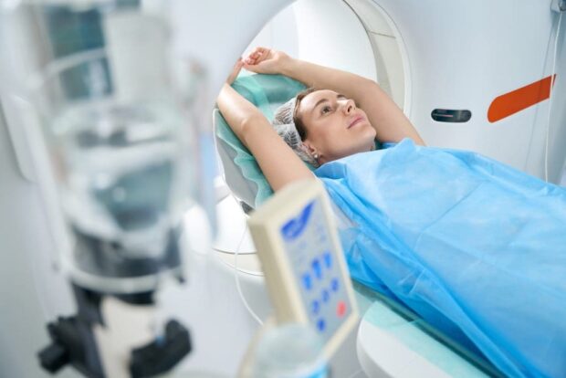
What to Expect During a Brain SPECT Procedure
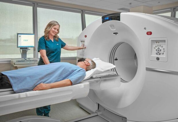
The brain SPECT imaging process is relatively straightforward and non-intrusive. Upon arrival at the imaging center, patients will first undergo a pre-procedure briefing, which includes a review of medical history and an explanation of the protocol. The procedure typically begins with the administration of a radioactive tracer via intravenous injection. After this, patients will be instructed to rest for a brief period to allow the tracer to circulate and target the brain.
After the waiting period, patients will lie on a table while a gamma camera captures images of their brain. This process usually takes between 20 to 60 minutes, during which patients are required to remain still to ensure high-quality images.
While the majority of patients experience no discomfort during the scan, some may feel a slight pinch during the injection of the tracer. After the procedure, there are no special precautions needed, as the radioactive substance will naturally diminish over time and be eliminated from the body. Most patients can return to their normal activities immediately following the SPECT scan.
Clarifying the Myths ─ Common Misunderstandings About Brain SPECT
Despite the demonstrated effectiveness and safety of SPECT imaging, several myths persist in public discourse. One common misconception is that brain SPECT scans are only useful for detecting severe neurological conditions. In reality, SPECT plays a critical role in identifying a broad spectrum of brain disorders and can also be instrumental in monitoring responses to treatment in lighter cases, not just severe ones.
Another misunderstanding revolves around the diagnostic usefulness of SPECT scans. Some people incorrectly believe that scans can provide a definitive diagnosis on their own. While SPECT provides valuable functional data, it is essential to integrate these findings with clinical assessments, patient histories, and other investigative modalities.
Diagnosing brain health conditions is a multifaceted process that requires a comprehensive approach. Thus, it is critical for patients to engage in an ongoing dialogue with their healthcare providers to interpret the results adequately and formulate appropriate treatment plans.